Stanford Medicine researchers have linked a specific gene known to be associated with ALS with a characteristic of the disease, opening avenues for a targeted therapy.
February 23, 2022 - By Hanae Armitage
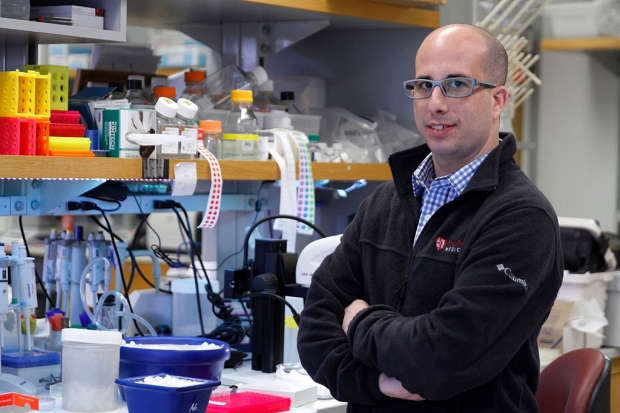
Aaron Gitler and his colleagues have found a connection between a genetic mutation often found in ALS patients and a prominent characteristic of the disease.
Paul Sakuma
Scientists who study amyotrophic lateral sclerosis know there are certain telltale features of the disease, and they occasionally find suspicious genes that seem to be connected to the development of ALS — but the two rarely align.
Now, a study led by researchers at Stanford Medicine and the Mayo Clinic has confirmed a connection between ALS’s most prominent molecular characteristic, a type of protein aggregate in the brain, and a gene that’s long been thought to contribute to the disease.
“We know that in about 98% of ALS cases, this erroneous protein aggregation forms,” said Aaron Gitler, PhD, a Stanford Medicine professor of genetics. “And past genetic studies show that there’s a gene, UNC13A, that’s obviously connected to ALS. It’s on everyone’s radar, but no one knows how it contributes to the disease. This finding connects the two — the most common pathology with one of the most common genetic risk factors.”
ALS, also known as Lou Gehrig’s disease, is a neurodegenerative condition that degrades nerve cell function and robs an individual of voluntary muscle movements. Walking, getting dressed or feeding oneself become virtually impossible as the disease progresses.
The discovery by Gitler and his group fills a big gap in the understanding of how ALS develops and could pave the way for a new treatment for the disease, which has no cure or effective therapies.
A paper describing the study published Feb. 23 in Nature. Gitler is the senior author. Rosa Ma, a graduate student in Gitler’s lab; Mercedes Prudencio, PhD, an assistant professor of neuroscience at the Mayo Clinic; and Yuka Koike, PhD, a postdoctoral scholar at the Mayo Clinic, share lead authorship.
Bad clumps
Neurons that exhibit clear signs of ALS often show the aggregation of a protein, called TDP43, outside its proper cellular home. Normally, TDP43 lives in the nucleus of a cell, where genes that encode the instructions to make a cell’s proteins are located. But in ALS, as the result of a poorly understood molecular glitch, TDP43 clumps outside the nucleus.
TDP43 is a crucial part of the protein-making process. Genes provide a template for proteins, but that genetic information needs to be preened and prepped before it can become a protein. That’s where TDP43 comes in: It engages in a quality-control-type role known as splicing, which cuts out superfluous molecular junk that can muck up an otherwise pristine protein.
In ALS, when TDP43 is missing from the nucleus and ejected into the cytoplasm — a sort of gel that bathes the cell’s other machinery — splicing doesn’t always happen correctly. Instead, the genetic filler that’s usually cut remains in some protein templates, creating something called cryptic exons, which, in the case of ALS, are as nefarious as they sound.
Where does UNC13A fit in? When TDP43 is missing from the nucleus of a cell, it can no longer cut out the unnecessary scraps of genetic information in preparation for protein production, leading to cryptic exons. The resulting protein can have diminished function, and this is what happens with UNC13A. (Think of TDP43 as a delete button that edits typos out of a sentence, with that sentence being a long sequence of molecular information coding for the UNC13A protein.) Just like typos make sentences hard to read, these cryptic exons interfere with the cell’s ability to read the genetic instructions to make the proper proteins.
It’s important to note that the mutation in UNC13A, in itself, is not a cause of ALS. If TDP43 is present, like it is in a healthy cell, the pernicious snippet can be excised right out of the protein, Gitler said. ALS results from both a mutation in UNC13A and a dysfunctional TDP43, among other causes. In that sense, UNC13A is a “risk factor,” meaning that the mutation in UNC13A can help predict a person’s risk for developing the disease.
“We think of it as an Achilles’ heel, a potential weak point that could lead to a neuron’s downfall,” Gitler said. He suspects there could be more of these “Achilles’ heels,” which could increase a person’s risk of developing ALS, lurking throughout the genome. And now, thanks to Ma’s new approach to genomic data analysis and subsequent experimental tactics, the team has a reliable way to find them.
Looking for cryptic exons
Past studies of the genetics of ALS have shown that UNC13A is commonly mutated in patients with the condition.
“Rosa Ma did something very clever — she reanalyzed the data from these past studies, but instead of looking for changes in gene expression, she looked for cryptic exons,” Gitler said. UNC13A immediately jumped out to Ma.
As it turned out, the UNC13A mutations that were flagged in the original genetic studies were occurring exactly in the same cryptic exon in UNC13A that Ma discovered — a detail previous studies hadn’t been able to show. Paired with the fact that TDP43 is known to be dysfunctional in ALS patients, the discovery sheds light on how TDP43 and UNC13A are connected: With TDP43 out in the cytoplasm, cryptic exons in UNC13A pop up, and within those cryptic exons, the mutated instructions that drum up neurological trouble.
Healthy UNC13A helps neurons signal to one another, a process that controls everything from memory to muscle control.
Through experiments on neurons in a lab dish, the researchers confirmed removing TDP43 from the nucleus causes cryptic exons to form in UNC13A. The cryptic exons in turn, decreased the function of the protein.
“We’re still figuring out exactly what this means, but we hypothesize that it’s basically communication between neurons getting shut down — and that’s really bad,” Gitler said.
Drug target
The next step is to see if UNC13A cryptic exons can somehow be muted — or their formation blocked — before cell communication is hindered. That’s what Gitler and his colleagues are working on now. Now that they know precisely where the ALS-linked cryptic exon in UNC13A is hiding, they are well on the way to designing a therapeutic strategy to keep it from popping up even when TDP43 causes problems. In addition, results from Ma’s genetic reanalysis pointed to cryptic exons in other genes, and Gitler’s lab plans to study them.
Although the new finding is a big step in connecting two widely known factors in ALS, Gitler cautions that this is not the sole mechanism of ALS — there are still more mysteries to solve about how it develops and how to best treat it.
Other Stanford co-authors of the study are graduate student Garam Kim; visiting graduate student Adam Briner; and postdoctoral scholars Caiwei Guo, PhD, Caitlin Rodriguez, PhD, Tetsuya Akiyama, MD, PhD, and Broder Schmidt, PhD.
Researchers from the Mayo Clinic, UC San Francisco, Maze Therapeutics, Johns Hopkins University, the University of Pennsylvania and the University of Queensland contributed to this study.
This study was funded by the National Institutes of Health (grants R35NS097263, R35NS097273, U54NS123743, P01NS084974, R01NS104437, RF1NS120992, RF1AG071326, RF1NS113636, P30AG06267 and U01AG006786), the Robert Packard Center for ALS Research at Johns Hopkins, the Brain Rejuvenation Project of the Wu Tsai Neurosciences Institute, Target ALS, Amyotrophic Lateral Sclerosis Association, and the Office of the Assistant Secretary of Defense for Health Affairs through the Amyotrophic Lateral Sclerosis Research Program (W81XWH-20-1-0242).
-
 Hanae ArmitageHanae Armitage is a science writer in the Office of Communications. Email her at harmitag@stanford.edu.
About Stanford Medicine
Stanford Medicine is an integrated academic health system comprising the Stanford School of Medicine and adult and pediatric health care delivery systems. Together, they harness the full potential of biomedicine through collaborative research, education and clinical care for patients. For more information, please visit med.stanford.edu.