New technology combines radiotherapy with real-time detection of cancer cells to target moving tumors or multiple metastases. Stanford Medicine is the first to research the technology in the clinic.
August 24, 2023 - By Krista Conger
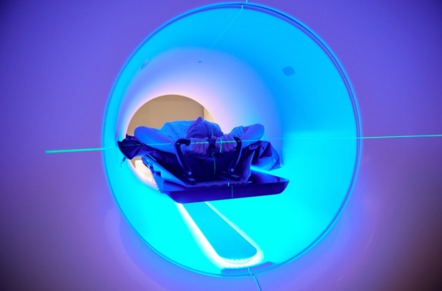
Stanford Medicine physicians are testing a machine that targets radiation based on real-time feedback from cancer cells.
RefleXion
Radiation therapy is a key component of care for many types of cancers. But some tumors can be more difficult than others to treat. Cancers in the lungs, for example, move with each breath, while tumors that have metastasized to many places in the body can require repeated radiation sessions.
Earlier this week, Stanford Medicine launched a new method of delivering radiation that uses signals from cancer-targeting molecules called tracers to target tumors in real time. It is the first time the new approach, known as biology-guided radiation therapy or SCINTIXTM, has been used in a clinic.
“This is the first radiation treatment machine in the world to combine radiotherapy with PET [positron emission tomography] technology,” said Michael Gensheimer, MD, clinical associate professor of radiation oncology. “It targets the cancer directly in areas where it is most active, tracking its movement and adjusting the radiation delivery several times a second.”
Although more real-world testing needs to be done, the goal is that the technology will eventually make radiation treatment faster and more precise, improving patient comfort, minimizing side effects and killing cancer cells more efficiently.
The machine, X1, was developed at Hayward, California-based RefleXion Medical in collaboration with clinicians and medical physicists at Stanford Medicine. In February, the Food and Drug Administration approved the platform for the treatment of certain tumors in the lungs and bone, based on a clinical trial at Stanford Medicine that simulated treatment delivery to patients.
“In addition to the clinical implementation and feedback leading to the first FDA-cleared treatment, the medical physics team worked tirelessly to validate the accurate delivery of the first treatment and ensure that the new technique was working as promised,” said professor of radiation oncology Billy Loo, MD, PhD. “It took a great team effort between physicians, the medical physics and dosimetry team led by Drs. Murat Surucu, Nataliya Kovalchuk and Bin Han, nurses, coordinators, and clinic staff to bring this milestone to fruition.”
A novel approach to radiation
“It is basically a new way to deliver radiation therapy,” said Lucas Vitzthum, MD, clinical assistant professor of radiation oncology. “We are excited to develop the technology and evaluate whether and how it might benefit our patients.”
Vitzthum is heading a clinical trial to assess the effect of the technology on clinical outcomes and quality-of-life measures for patients, as well as any possible adverse side effects.
The real-time feedback sets the technology apart from the way radiation is delivered now. Currently, cancers are mapped in the body using small molecules called PET tracers. These tracers are given to the patient intravenously. One part of the tracer is designed to bind to cancer cells; the other emits a signal that can be detected outside the body. A PET machine detects where in the body the signals, and therefore the cancer cells, are located. PET scans are then used to plan which areas of the body to irradiate.
“Standard radiation therapy treatment plans are generally tailored to static 3D representations of the tumor and normal anatomy taken days to weeks before treatment,” Vitzthum said, noting that anatomy can shift over time. “In contrast, the PET-guided radiation therapy machine is a closed-loop system — continuously detecting PET signals in real time during radiation treatment. This has the potential to help us precisely target the radiation and spare healthy cells.”
If the promise is realized, biology-guided radiation therapy may be a boon to patients with tough-to-treat cancers. Vitzthum notes that it will be important to compare this technology with existing treatment approaches using prospective randomized controlled trials.
“I once had a patient with early-stage lung cancer whose breathing was very irregular, making it difficult to effectively irradiate the tumor even with long treatment sessions,” Gensheimer recalled. “We ended up implanting metal markers into the tumor to allow us to safely deliver treatment. But this was an invasive approach that required IV sedation. Our hope is that this technology will help us better treat patients like this one less invasively.”
Biology-guided radiation therapy may also assist patients with advanced disease who currently have fewer treatment options.
“We plan to use this technology to explore the role of radiation in metastatic disease,” Vitzthum said. “It may eventually be possible to treat multiple tumors in the body simultaneously.”
-
 Krista CongerKrista Conger is a senior science writer in the Office of Communications. Email her at kristac@stanford.edu.
About Stanford Medicine
Stanford Medicine is an integrated academic health system comprising the Stanford School of Medicine and adult and pediatric health care delivery systems. Together, they harness the full potential of biomedicine through collaborative research, education and clinical care for patients. For more information, please visit med.stanford.edu.