Combining two cutting-edge technologies, researchers revealed the impact of a multitude of genes that are associated with neurodevelopmental disorders, including autism, but whose effects on human brain development were previously unknown.
September 27, 2023 - By Bruce Goldman
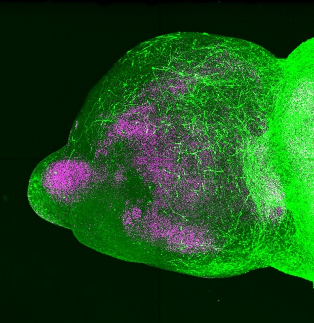
Forebrain assembloids: The human migrating interneurons are in green; nuclei are in pink.
Pasca lab
Stanford Medicine investigators and their colleagues sifted through a jumble of genes implicated in neurodevelopmental disorders and identified dozens of disparate troublemakers with similar effects.
Because the method they used sorts defective genes by their function — or in this case, their dysfunction — the approach is likely to accelerate drug development for neurodevelopmental disorders.
Studies have implicated at least 500 genes in such disorders. But scientists have no idea exactly how defects in most of these genes impair brain function.
The research, which points the way toward creating order from this chaos, is described in a paper published online Sept. 27 in Nature. Sergiu Pasca, MD, the Kenneth T. Norris, Jr. Professor II of Psychiatry and Behavioral Sciences, is the study’s senior author. The lead author is Xiangling Meng, PhD, a postdoctoral scholar in Pasca’s group.
There are two main classes of neurons in the cerebral cortex: excitatory and inhibitory. Excitatory neurons fire impulses that activate other neurons, while inhibitory neurons’ firing blocks other neurons from firing. Inhibitory and excitatory neurons integrate to form circuits, shaping signaling activity in the brain.
In humans, as many as half of all cells in the cerebral cortex, the brain’s outermost and most recently evolved layer, are inhibitory. Scientists theorize that an imbalance in the number or function of inhibitory neurons compared with excitatory neurons might be at least partly responsible for autism spectrum disorder and epilepsy.
“If that’s true, you could find ways to alter the functional balance of these cells in the cortex as a therapeutic approach for these disorders,” Pasca said. But first, he wondered, how do you make sense of the enormous collection of genes that have been implicated in these conditions but whose impacts are largely unknown?
Does the existence of hundreds of genes associated with disease mean that there are hundreds of different types of neurodevelopmental disorders, each requiring a different remedy? Or might several different genes converge and lead to similar pathology, or some particular aspect of it?
“If it’s the latter, a cluster of gene defects that all produce a similar physiological deficit might be amenable to a single type of treatment,” Pasca said.
A new avenue of research
Until recently, there was no way to study early brain development in a human being. But Pasca, who is also the Bonnie Uytengsu and Family Director of the Stanford Brain Organogenesis Program, pioneered a technology that makes detailed exploration of the developing human brain possible. With special laboratory glassware; a combination of growth factors (substances that stimulate cell growth) and nutrients; and human induced pluripotent stem cells — or iPS cells, which can be generated from a simple skin biopsy — he can generate small clumps of neural tissue whose anatomical architecture and function closely resemble part of the brain, such as the human cerebral cortex. Pasca calls these clumps cortical organoids.

Sergiu Pasca
Adjusting the nutrients and growth factors in which iPS cells are bathed, Pasca has shown, generates organoids approximating another brain structure called the subpallium. Located deeper in the forebrain than the cortex, the subpallium plays a key role during fetal and infantile development: It produces inhibitory neurons, known as interneurons, that migrate to the cortex and elsewhere and join up with excitatory neurons to form functioning circuits capable of complex signal generation.
By placing subpallial organoids next to cortical organoids and waiting for several days, Pasca was able to observe, for the first time, how these two structures fuse together to form what he calls “assembloids” and to watch interneurons migrate from the subpallium to the cortex and hook up with excitatory neurons there.
In the new study, Pasca and his colleagues paired this cutting-edge technology with another: CRISPR.
CRISPR employs a molecular scissors and a molecular bloodhound. This enables researchers to snip out specific DNA sequences at will, so they can knock selected genes out of the genome and see what happens.
Pasca and his colleagues narrowed the neurodevelopmental-disorder-associated genes on their list down to 425 that are activated in interneurons. They created iPS cells that when differentiated into multi-celled organoids fluoresced only in those cells that had become interneurons. This way, they could generate subpallial organoids in which they’d be able to distinguish interneurons from all the other brain-cell types the subpallium produces.
They infected thousands of these cells — which had also been bioengineered to carry a gene whose protein product was the molecular scissors of CRISPR — with CRISPR “bloodhounds”: small strips of nucleic acids that are complementary to portions of one or another of the 425 neurodevelopmental-disorder-associated genes of interest. Wherever such a strip affixed itself to a gene, the CRISPR “scissors,” an enzyme, would excise the DNA bound by the strip.
Tossing enough of these bloodhounds into a large enough pool of these cells made it statistically likely that each of the 425 suspect genes would get snipped out in at least hundreds of cells in the pool.
From those iPS cells, the researchers generated subpallial organoids. Some showed no fluorescence at all after 44 days in a dish, indicating their failure to generate interneurons. By sequencing the genomes of these organoids, the researchers could tell which gene had been disabled by the CRISPR scissors, resulting in defective interneuron generation. They identified 13 such genes.
To hunt for genes affecting interneuron migration, Pasca’s team placed subpallial organoids capable of generating interneurons beside healthy cortical organoids in numerous lab dishes. The two types of organoids in each dish fused to form assembloids. After about 30 days of organoid cohabitation, the researchers pulled out about 1,000 assembloids from different dishes and cut each of them apart at the fusion junction. Then they compared the fluorescing interneurons in each assembloid’s subpallial section with those that had crossed the finish line into that assembloid’s cortical section.
By sequencing the genomes of cells, the scientists could pinpoint genes in which a targeted deletion had impaired migration. They found 33 of them.
“The identification of 46 genes — close to 10% of all known neurodevelopmental-disorder-associated genes — whose dysfunction impairs interneuron development points to a subgroup of neurodevelopmental disorders that are characterized by inadequate inhibition of excitatory cortical neurons,” Pasca said.
Some of these genes have been previously uncharacterized, and their function unsuspected, Pasca said.
But other genes have been identified in other studies. “This was a comfort factor,” Pasca said. “It meant we weren’t insane — this approach works.”
Researchers from the University of California, San Francisco, contributed to the work.
The study was funded by the Stanford Brain Organogenesis Program in the Wu Tsai Neuroscience institute and Bio-X, Kwan Funds, Senkut Funds, the Ludwig Foundation, the Mann Foundation, the Chan Zuckerberg Initiative, CZ BioHub, and the New York Stem Cell Foundation.
-
 Bruce GoldmanBruce Goldman is a senior science writer in the Office of Communications. Email him at goldmanb@stanford.edu.
About Stanford Medicine
Stanford Medicine is an integrated academic health system comprising the Stanford School of Medicine and adult and pediatric health care delivery systems. Together, they harness the full potential of biomedicine through collaborative research, education and clinical care for patients. For more information, please visit med.stanford.edu.